GLOSSARY (ticks)
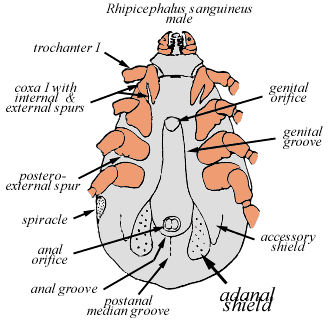
adanal shields: Also called adanal plates, these are posteriorly directed sclerotised plates on the ventral surface near (usually posterior) to the anus in males of some ixodid ticks.
alloscutum: in Ixodid ticks, the dorsal body region posterior to the scutum. When examined under electron microscope , this region shows numerous fine striations representing superficial folds of the cuticular surface. In the mid-dorsal region are the foveae dorsalis (=foveal pores), paired structures containing numerous tiny pores. These structures are most evident in females, but may also occur in males (absent in the genus Ixodes). These structures have a role in pheromone secretion and mating.
anterior projection: anterior extension of the body beyond the capitulum in argasid ticks, so that the capitulum is invisible from the dorsal aspect
anal aperture/anus: the posterior terminus of the alimentary canal. The aperture is bordered by the anal plates and surrounded by a cuticular ring in some ixodid species. The anus is situated in the median line posterio to the coxae. The term applies to the external the external anal apparatus consisting of a more or less evident ring within which are two laterally moving valves.
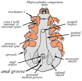
anal groove: in hard (Ixodid)
ticks, a groove adjacent to the anus. In ticks of the genus Ixodes
(Prostriata), the anal groove originates on or near the
posterior body margin and surrounds the anus anteriorly. In most
cases the grooves fuse in a semicircle in front of the anus, in
others they form an ogive (hence ogival, like a gothic
arch). In some species the groove are not continuous anteriorly.
In other hard ticks (Metastriata) the anal groove is
smaller and lies posterior to the anus. In most cases they run
forward and outward toward the genital grooves, which they may
attain. In some cases they are continuous with a postero-median
groove from which they fork anteriorly.
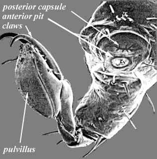
apotele: Also called the pre-tasus. The terminal segment of the tarsus made up of the claws and a pad (pulvillus). The pulvillus is virtually absent in argasid nymphs and adults.
articles: Segments of the legs or palps. Usually reserved for the palps. Article 1 is adjacent (proximal) to the body. Ixodid ticks have four articles, the fourth beng recessed in the apex of article 3.
auriculae: paired extensions of the ventral posterior or lateral margins of the basis capituli.
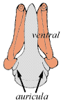
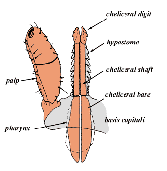
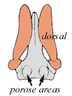
basis capituli: Also called the "basal ring", or "Kragen". The basal ring of cuticle to which the palps, chelicerae and hypostome are attached. The basis capituli may be moved in the dorsoventral plane, and articulates with the body proper. It often shows a transvers elevated "dorsal ridge" with edge directed backward. The ridge may have protruding angles, the latter termed "cornua". There may also be a "ventral ridge". The "auricula" signifies a protruding retrograde process at the lateral angles of the ventral ridge posterior to the insertion of the palps. When the length of the capitulum is given it is measured from the tip of the hypostome to the "dorsal ridge" in the median line. Where the dorsal ridge is absent it is similarly measured to the "ventral ridge". When the measurement is made ventrally this should be stated, the dorsal method being usually employed. The clear space between the porose areas (only present in the female capitulum) is spoken of as the "interval".
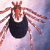
body: Also called the idiosoma. The entire body (scutum and alloscutum) is covered by numerous setae (hairs) and less evident pore-like sensilla. Most are ear-like in appearance (sensilla auriformia) but other types also occur. In larvae, relatively few setae occur, and their number and relative placement provide valuable characters for generic and sub-generic differentiation (chaetotaxy). The body of the paralysis tick contains the following visceral organs- midgut (along the midline), the midgut diverticulae (diverging laterally), the large paired salivary glands (situated laterally, see diagram of salivary gland), the rectal sac, the genital organs, the malpighian tubules, the tracheae and tracheal trunks (entering laterally) and the synganglion. See diagram of viscera.
camerostome: In argasid ticks, the cavity in which the capitulum is situated
capitulum: Also called the "rostrum", "the head", or "false head"and the "snout". It is the moveable anterior extension of the body (the idiosoma) and includes the palps and mouth parts. It is not a head in the sense that it does not contain the tick's "brain", nor the salivary glands, nor even eyes (where present)- all of these are located in the body of the tick. The capitulum articulates with the body via a cavity (the emargination in ixodids and the camerstome in argasids), and normally lies in the same plane as the body. It is connected by a soft articulation membrane that allows the capitulum to be flexed (ventrally) or extended (returned to the horizontal axis). See also palps, chelicerae, hypostome and basis capituli. The hypostome is separated from the chelicerae in ixodids by a membrane and in argasids by the labrum. Take a look at a close up view of the capitulum of Ixodes holocyclus. See anatomy for a more complete description of these structures.
cervical grooves: paired grooves on the scutum (shield) extending posteriorly from the inner angles of the scapulae (shoulders). May be shallow, deep, faint or absent.
cheeks: paired, movable flap-like covers orientated in the anterior plane and which can close medially, covering and protecting the capitulum in some species of Argasidae.
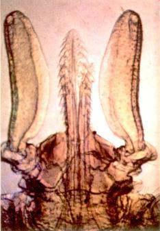
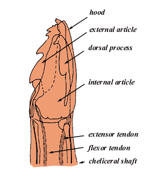
chelicerae: Also called "mandibles" or "pseudo-chelicerae". The first pair of appendages of ticks and other arachnids lying dorsal to the hypostome. The tick chelicerae consist of 3 segments, the cheliceral bases located within the basis capitulum, the cheliceral shafts that originate within the basis capituli and extend anteriorly, and the cheliceral digits that bear the denticles. The cheliceral shafts are surrounded by tough, spinous sheaths which are an outgrowth of the basis capituli. A delicate sheath (the cheliceral hood) covers the digits in most species.
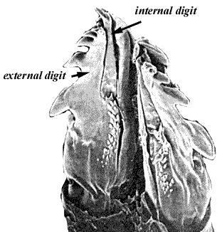
cheliceral digits (=cheliceral articles): the cheliceral digits are terminally appended to the cheliceral shafts. They lie dorsal to the hypostome and bear laterally directed cutting edges. Their movements result in ripping and tearing against the host skin. The internal (medial) digit is moveable, and is directed from side to side by tendons attached to the powerful muscle masses in the bulbous cheliceral bases (not externally visible). The internal digit lies within a cavity of the outer digit and the two move together.Both digits have sharp denticles. The inner digit bears mechanosensory and chemosensory receptors providing information about shear forces and chemical composition of host fluids. They also play a role in pheromone detection.
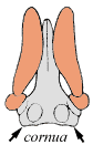
cornua: Paired posterior projections of the postero-lateral margins of the dorsum of the basis capituli in many ixodid ticks.
 |
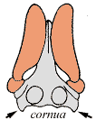 |
| the blunt cornua of Ixodes holocyclus | the sharp cornua of Ixodes cornuatus |
corona: a field of tiny denticles at the anterior end of the hypostome.
coxa: the basal segment of each leg that articulates with the body and to which body muscles attach. The coxae are only slightly movable. In the males of some species the coxae are greatly enlarged and cover most of the ventral surface of the body. When coxae are described as bifid they bear two spurs and are deeply incised. When descibed as trenchant they have a knife-like margin.Coxae are numbered I, II, III and IV from anterior to posterior.
crenelations: small hypostomal ridges or denticle-like structures found in some species, usually in non-feeding males; may also occur beyond the denticulate zone in species in which the hypostome bears prominent denticles.
denticles: the teeth or small, recurved projections on the ventral side of the hypostome and at the end of the cheliceral digits.
dentition: referring to the arrangement of denticles on the hypostome, eg 2/2 refers to 2 longitudinal files of denticles on either side of the miudline of the hypostome.
discs: flattened depressions, usually subcircular, with a smooth or mottled surface, representing the sites of dorso-ventral muscle attachments.
dorsal humps: prominent elevations on the dorsal surfaces of certain leg segments, not present on all species.
dorsal plate: a smooth surface, usually elongated or subcircular, on the dorsum of the larvae in many argasid ticks; not to be confused with the scutum of ixodid ticks.
ecdysis: sloughing or casting off of the outer epidermis, desquamation.
emargination: in ixodid ticks the body cavity in which the basis capituli is situated (synonymous with camerostome).
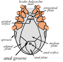
epimeral plates: paired ventral plates in males of the genus Ixodes, situated laterally and posteriorly to the spiracular plates.
eschar: a dry slough, especially that produced by heat or a corrosive or caustic substance
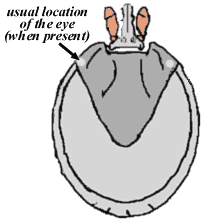
eyes: found on the scutum of certain ixodid ticks, namely Boophilus sp, Margaropus sp, Rhipicephalus sp, Amblyomma sp, Dermacentor sp, and Hyalomma sp, but NOT Haemaphysalis sp and Ixodes sp. Whilst many tick species lack eyes, nevertheless all ticks studied hae been found to have some type of photosensilla and also have optic ganglia in their synganglion. In hard ticks with eyes, paired simple eyes are located on the lateral margins of the scutum. In soft ticks possessing eyes, they occur on the ventrolateral surface, lateral to the legs. The eye is similar in all 3 active life stages. Little is known about the physiology of vision in ticks but based on the morphologic descriptions, it is extremely doubtful that the eyes are capable of detailed form perception. Ticks with well developed eyes seem to respond to shadows, variations in light intensity and perhaps are capable of vague discrimination of shapes. When questing D. variabilis responds to shadows by extending its legs, ready to attach to a host (Sonenshine, 1991).
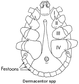
festoons: in some ixodid ticks; small portions of the ventral posterior body margin, marked by delicate grooves, usually rectangular.
folds: prominent cuticular ridges on the ventral or marginal surface of the body in argasid ticks.
Gene's organ: a structure on the female tick's dorsum, between the basis capituli and the scutum (the camerostomal cavity of argasids, or emargination cavity of ixodids) producing a wax which coats the eggs.
genital aperture: is located in the approximate midline between the coxae. In females the genital aperture appears as a posteriorly directed U- or V-shaped groove, with prominent marginal folds. The shape of the female genital pore varies greatly among the species and provides useful characters for taxonomic study. In males of the Metastriata, the genital pore is covered by by a moveable plate which can be elevated when a spermatophore is to be passed
goblets: tiny cavities within the cuticle of the spiracular plate, appearing as subcircular structures when viewed from the external surface.
granulations: irregular bumps or elevations on the integumental surface. See punctations
grooves: deep, linear depressions in the body cuticle, usually on the ventral surface.
Haller's organ: a sensory structure on the dorsal surface of the tarsus of leg1. It is comprised of an anterior pit and a posterior capsule. It is present in all post-embryonic stages of all species of ticks. It is a complex sensory apparatus that is very important as the primary organ for determining host location, host odours, detecting pheromones and other sensory functions.
hood: in argasid ticks, the anterior projection of the camerostome, anterior to the capitulum (not to be confused with the cheliceral digit hood)
hypostome: the median ventral extension of the basis capituli, between the palps and the chelicerae, and covered with recurved teeth on its ventral surface; with a pronounced pre-oral canal (the hypostomal gutter) on its dorsal surface. See anatomy for a diagram. Also called "maxilla", "radula", "labium" and "Unterkiefer". The dentition is indicated by figures on either side of a vertical line. Thus 3|3 means 3 longitudinal files of teeth on each half of the hypostome. The hypostome may be pointed, rounded or emarginated distally. It may be armed from tip to base with teeth or only bear teeth along a part of its length. An unarmed, protruding median ridge, which broadens basally, may run down the length of the hypostome, starting near the tip. When a hypostome is described as having a corona, the tip bears a number of very minute denticles.
imago: an adult sexually mature insect or acarid produced after metamorphosis [C18: New Latin, from Latin: likeness]
instar: the stage in the development of an insect or acarid between any two moults [C19: New Latin, from Latin: image]
Ix*o*des \"ik-'sO-(")dEz\ n : a widespread genus of ixodid ("hard") ticks many of which are bloodsucking parasites of humans and animals, and sometimes cause paralysis or other severe reactions
ixodiasis: a disease caused by or transmitted by ticks. From New Latin Ixodes [The Chambers Dictionary. 1994 by Chambers Harrap Publishers, Ltd.]
ixos: Gk= mistletoe. Is there a connection with the tick terminology "ixodes"?
lateral carinae: ridges on the lateral margins of the scutum in some ixodid species.
legs: For ticks the first pair of limbs
form the chelicerae (=mandibles) for cutting into the skin
of the host, whilst the second pair of limbs forms the palps
which position the capitulum for feeding. The remaining four
pairs of limbs form the actual legs.
In ticks the legs are used for walking, both on and off the host.
The four pairs of legs are each divided into 6 segments (articles)-
coxa, trochanter, femur, tibia, metatarsus and tarsus. The coxae are situated on the ventral side of
the body and have very limted movement and support the articles.
The articles are connected by a soft articulation cuticle, seen
readily when the artciles are flexed. In some species the femur
appears superficially divided into a basifemur and a telofemur
but this division is not reflected in the musculature and these
subdivisions are not moveable. A similar subdivision may also be
noted in the tarsus
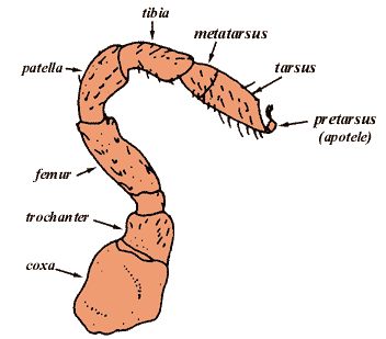
Of the articles the first (trochanter) and last (tarsus) are of importance in classification. All of these may bear spurs, spines or teeth in various species. It is important to note the manner in which the tarsi terminate :if they taper, are humped, bear ventral spurs etc. The length of the claws in relation to the pads, or suckers (pulvillum) should also be noted. The other segments are highly flexible but are mainly used by flexing and extending the joints. They may be folded against the ventral body wall for protection. The segments are connected by a soft articulation cuticle. The tarsus of each leg bears an apotele (=pre-tarsus), including the claws and and the pulvillus. There is a complex sensory apparatus (=Hallers organ) located on the dorsal surface of the tarsus of leg 1. primary organ for determining host location, and detecting host odors and pheromones etc.
macula: a prominent sclerotised fold or hump constituting part of the spiracular plate.
mammillae: minute conical or truncated integumental elevations covering the body surface and legs in Ornithodoros ticks; distinct from tubercles or granulations found in other argasid species.
marginal groove: in Ixodes a groove situated along the dorsal marginal surface of the body, most evident in females.
mandibles: see chelicerae
median plate: in males of the genus Ixodes the sclerotised plate between the coxae, posterior to the genital orifice, on the ventral side of the body.
ornamentation: having symmetrical enamel-like colour patterns in the sclerotised (hardened) cuticle, usually iridescent, and confined to the scutum and/or the dorsal surface of the basis capituli and legs of some ixodid ticks. Ticks on which such ornamentation occurs are spoken of as "ornate". For example Dermacentor spp and Amblyomma spp.
palps: the second pair of appendages, normally with four segments termed articles; article 1 articulates with the basis capituli; in argasids article1 is elongated, about as long as articles 2 and 3, and movable; in ixodids, article 1 is usually immobile and shorter than articles 2 and 3; articles 2 and 3 varying length, are almost always the longest segments of the palps, and are more flexible; article 4 is terminal in argasid ticks, but recessed in a cavity on the ventral side of article 3 in most ixodid ticks. This delicate segment can be retracted or protruded as needed. Numerous fine hair-like structures (setae) presumed to function as chemosensilla, occur on the tip of this segment and form a sensory field. Other setae, often long and stout, occur on the ventral and medial surfaces of the palps and serve to shield the delicate mouthparts.
plates: are large dense armour-like chitinous structures occuring in Ixodes males, not rising above the surfaceof the body (as do the shields for instance in Rhipicephalus etc). The plates are bounded by the ventral grooves or by soft portions of the integument. There are 1 pregential, 1 median and 1 anal plate along the median line of the body; there are 2 adanal plates to either side of the anal plate and 2 epimeral plates with indistinct external borders extending forward outside the genital grooveto near coxa IV.
porose areas: clusters of tiny depressions on the dorsal surface of the basis capituli in ixodid females. In some species the porose areas are large and cover almost the entire dorsal surface.
pulvillus: a pad-like structure on the terminal end of the tarsi, adjacent to the claws in ixodid ticks; also present in the larvae of most argasid ticks.
punctations: are circular depressions dotting the integument, and frequently bearing hairs. Those parts on which punctations occur (scutum, alloscutum, capitulum etc) are referred to as finely or coarsely punctate. The punctations mostly represent sensory organs (sensilla) some of which contain hair-like sensory structures (setae).
replete: adj. (usually postpositive) 1. (often foll. by with) copiously supplied (with); abounding (in). 2. having one's appetite completely or excessively satisfied by food and drink; stuffed; gorged; satiated [C14: from Latin repletus, from replere to refill, from RE- + plere to fill].
Rickettsia: genus of bacteria , named after the American pathologist Howard Taylor Ricketts, who first discovered them. Like viruses , they can survive and multiply only inside cells . The micro-organisms, which are either round or long, are found in mammals and cause several diseases in human beings. They are generally carried by arthropods, such as fleas, lice, mites, and ticks . Numerous insect-infesting species of Rickettsia do not cause diseases in mammals. Rickettsial diseases include different forms of typhus, Rocky Mountain spotted fever, and trench fever. Q fever and rickettsial pox are also rickettsial diseases. Rickettsial diseases are treated with antibiotics and prevented by destroying the arthropod carriers (Concise Encarta, 1999, Microsoft).
salivarium: the combined part of the salivary duct located within the capitulum.
scapulae: shoulder-like projections on the antero-lateral edges of the scutum in many ixodid ticks.
scutum: the dorsal sclerotised plate (shield) covering the anterior part of the body in female ixodid ticks (the entire dorsal surface in males); also called the "face"; not to be confused with the dorsal plate on the body of the larvae in most argasid species. Macroscopically. the scutum may bear distinct grooves, the cervical grooves, and ridges, the lateral carinae. In some species the lateral margin of the scutum bears the paired eyes. In Hyalomma spp, however, prominent eyes occur on the softer cuticle adjacent to the scutum. In females, a paired protrusible organ (Gene's organ) may be visible in the camerostomal or emargination cavity between the scutum and the basis capituli. The scutum is smooth in most species, but highly punctate or rugose patterns may also occur.
sensilla: plural the sensory organs for monitorig the status of the internal and external environment in insects and ticks. Functionally there are several types- chemosensilla, mechanosensilla, photosensilla and thermosensilla. Combinations of these, multifunctional sensilla, also occur . Other sensilla with hygroreceptors and even osmoreceptors have also been postulated but not confirmed. Many sensilla are diffusely dispersed over the body and appendages. Others are clustered in specialised organs such as the Haller's organ (on the foreleg tarsi), the palpal organ on palpal article IV, the organ on the cheliceral digits, the eyes and so on.
setae: plural of seta, (in invertebrates and some plants) any bristle or bristle-like appendage [C18: from Latin].
setaceous: bristle like
shields: These are salient chitinous structures occuring in the males of Rhipicephalus, Boophilus and Hyalomma on either side of the anus. A pair close to the anus is always present and is termed adanal. There is frequently a second pair external to these, known as accessory, these being well-developed in Boophilus.
spiracle: Also called "spiracular plate" or "peritreme (gamasid mites)" or "stigmal plate". The spiracular plate and the macula together form the spiracle. The spiracle is a respiratory organ situated ventrolaterally posterior to coxa IV. It may be circular, oval or comma-shaped. In all ixodid ticks the spiracular plate is and shows a more or less central structure called the macula adjacent to the small often slit-like ostium which is the opening to the respiratory sysytem. The punctate surface represents the semi-transparent goblets, which in turn represent internal spaces (aeropyles). The number and arrangement of these goblets vary greatly and occasionally provide useful characters for species separation within the Ixodidae. The spiracular plate may be enclosed by a more or less broad frame of chitin which is incomplete postero-dorsally in comma-shaped forms. The spiracles open into the atrium, a large sinus-like structure from which the tracheal trunks arise. The structure of the spiracle is useful in classification.
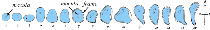
This figure (Nuttall and Warburtin, 1908) illustrates the spiracles of 15 species of Ixodidae to sjow the different forms they may assume. A= anterior, V= ventral, D= dorsal. 1- round as in I. ricinus female; 2- bluntly as in I. boliviensis female, 3-elongate sub-oval as in I. tasmani female; (in both 2 and 3 long axis of spiracle transverse to body axis); 4- elongate oval as in I. angustus female; ovoid flattened posteriorly (or sub-triangular) as in I.angustus male (in 3,4,5 long axis normal, ie directed forward); 6 with slight postero-dorsal protrusion as in Haemaphysalis hystricis male; 7- bluntly ovoid as in Boophilus annulatus female; 8- short comma-shaped as in Dermacentor variabilis female; 9- short comma-shaped as in Amblyomma longirostrum female, with frame broadened dorsally; 10- sub-triangular, with rounded angles and with frame broadening much dorsally as in Amblyomma geomyedae female (7-15: with distinct darkly-chitinised marginal frame "complete" in 7, incomplete posetero-dorsally in the remaining spiracles which are comma-shaped in 10, sub-triangular with rounded angles;11- Rhipicentor nuttalli male; 12- Dermacentor reticulatus male and Rhipicentor bicornis male; 13- Rhipicephalus sanguineus male and Amblyomoma versicolor male; 14- Dermacentor andersoni male; 15- Hyalomma aegyptium male and Rhipicephalus sp male (11-15: more or less comma-shaped spiracles). The macula is central in 1, eccentric in the others, being situated ventrally and anteriorly, and possessing a variable form.
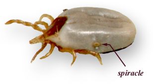
spiracular plate: the modified plate-like structure on the ventrolateral surface of the body caudal to coxae IV; in argasid ticks the plate consists of a very small simple elevated plate termed the macula and the spiracular opening next to an adjacent ridge of cuticle, all on the supracoxal fold; in ixodid ticks, the spiracular plate is a prominent structure with numerous ovoid air spaces within the cuticle (goblets) which are visible externally and give this structure its distinctive appearance, and the distinct macula closely approximated by the ostium.
supracoxal fold: a prominent fold on the lateral ventral margin of the body in argasid ticks, lateral to the legs; this fold bears the spiracles and, when present, the eyes.
tarsus:
The terminal portion of the leg of the tick, supporting the apotele (=pre-tarsus). In the
following examples transverse line indicates the pseudo-articulation.
See also the leg.
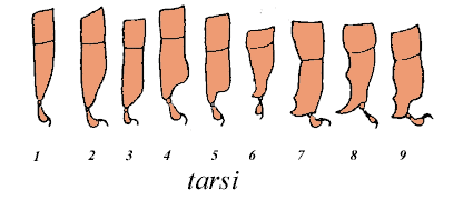
This diagram (Nuttall and Warburton, 1908), illustrates
the tarsi of 10 species of Ixodidae to show different forms of
tarsus 4:
1- tapering gradually as in I. ricinus; 2- tapering gradually as in I. signatus; 3- tapering obliquely as in I.ornithorhynci; 4- humped prior to tapeiring as in I. hexagonus; 5- tapering abruptly as in I. cordifer; 6- tapering to one spur as in Haemaphysalis cornigera; 7- ending bluntly with two spursas in Rhipicephalus masseyi; 8- tapering to a long spur as in Margaropus winthemi; 9- tapering abruptly and bearing two spurs as in Amblyomma cooperi.
tick: any of various small parasitic arachnids of the families Ixodidae (hard ticks) and Argasidae (soft ticks) typically living on the skin of warm blooded animals and feeding on the blood and tissues of their hosts; any of certain other arachnids of the order Acarina (mites and ticks); applied also to the sheep-ked and similar bloodsucking Diptera parasitic on cattle and horses, etc.[Old English ticca; Middle High German zeche tick; Middle Irish dega stag beetle].
trans-stadial: occuring across a stage or stages within a life cycle. For example, a disease organism that passes from larva to nymph to adult is one that is transmitted trans-stadially.
vector: an organism, esp. an insect or acarid, that carries a disease-producing microorganism from one host to another, either within or on the surface of its body [C18, from Latin: carrier, from vehere, to convey].
Bibliography:
Osol, A Ed. Blakistons Pocket Medical Dictionary, 3rd ed, McGraw-Hill Book Company, New York, 1973.
Nuttal GHF, Warburton C, Cooper WF, Robinson LE; Ticks, A Monograph of the Ixodoidea, Cambridge University Press, 1908.
Sonenshine, DE: Biology of Ticks, 2 volumes: Oxford University Press, New York, Oxford, 1991. (An excellent text on the general biology of ticks, available at Badham Library, University of Sydney)
The Paralysis Tick of Australia - Home
E-mail Us to report a broken link!