THE TICK LESION
An overview of tick feeding
The salivary glands
Attachment of the paralysis tick to it's
host
The host reaction
An overview of tick feeding
Ticks are obligate blood feeders. All active stages (larvae, nymphs and adults) require blood as a source of nutrition (except for a few argasid genera in which the adult mouthparts are non-functional, ie Antricola, Otobius and Nothoaspis). Adults also require the blood for sperm or egg production. The feeding process of ixodid ticks has first a slow phase for several days followed by a fast phase in the last 12-24 hours before detachment. There may be a ten fold increase in fed:unfed weights by the end of the slow phase, but there is an additional ten fold increase by the end of the final fast phase. Leaving the full engorgement as late as possible reduces the chances of detection and removal by the host . The process of feeding is called engorging. The hypostome has a groove along its dorsal surface forming a food canal (also known as the preoral canal) through which blood is drawn from the host and passed on to the mouth and pharynx. During blood feeding by ixodid ticks, the liquid portion of the meal is first concentrated by removal of water and excess ions, which move across the gut epithelium and enter the ticks body cavity (hemocoele). These components are then taken up by the salivary glands which produce a watery saliva that is injected back into the host (Cupp, 1991).
Blood meal digestion in ticks is similar in all species. The digestive system in both ixodid and argasid ticks is histologically divided into foregut, midgut and hindgut. The foregut comprises the sucking pharynx and the oesophagus. The midgut contains a ventriculus with a valve, a variable number of blind diverticula (caeca), and a rectal tube. The hindgut is divided into a rectal bulb and the rectum itself.
The Salivary Glands
Paired salivary glands are situated laterally in the body cavity of the tick (both argasid and ixodid). They comprise large clusters of acini. A central duct extends posteriorly for the length of the gland and anteriorly into the capitulum where the paired ducts fuse to form the salivarium (Sonenshine,1991)
Attachment of the Paralysis Tick to it's Host
Once it has chosen a feeding site a tick
positions itself with its legs so as to elevate its body at a
sharp angle. 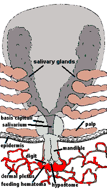 Guided
by the palps, the mandibular digits (chelicerae) cut into the
skin with their horizontal cutting action. These rip and tear at
the epidermal layers and a small pool of blood is formed. The
hypostome is inserted and this provides the initial attachment
strength. In the case of Ixodes holocyclus the hypostome
is inserted very deep into the dermis. The palps remain spread
apart on the surface. The process by which ixodid and argasid
ticks feed is termed telmophagy (=pool feeding). [This
contrasts with the process of solenophagy, used by
mosquitos, in which feeding is direct from a small venule.] The
resultant pool expands as a result of the anticoagulants released
from the salivary glands. In some Ixodid ticks a cement is
secreted into the wound within 5-30 minutes of cutting into the
skin. This material hardens quickly into a latex-like covering
around the mouthparts but excluding the palps that remain
flattened out on the skin surface. (Sonenshine, 1991). Ixodes
holocyclus, however, is one of the ixodid ticks which does
NOT produce cementum (Kemp, Stone and Binnington, 1982).
Guided
by the palps, the mandibular digits (chelicerae) cut into the
skin with their horizontal cutting action. These rip and tear at
the epidermal layers and a small pool of blood is formed. The
hypostome is inserted and this provides the initial attachment
strength. In the case of Ixodes holocyclus the hypostome
is inserted very deep into the dermis. The palps remain spread
apart on the surface. The process by which ixodid and argasid
ticks feed is termed telmophagy (=pool feeding). [This
contrasts with the process of solenophagy, used by
mosquitos, in which feeding is direct from a small venule.] The
resultant pool expands as a result of the anticoagulants released
from the salivary glands. In some Ixodid ticks a cement is
secreted into the wound within 5-30 minutes of cutting into the
skin. This material hardens quickly into a latex-like covering
around the mouthparts but excluding the palps that remain
flattened out on the skin surface. (Sonenshine, 1991). Ixodes
holocyclus, however, is one of the ixodid ticks which does
NOT produce cementum (Kemp, Stone and Binnington, 1982).
The Host Reaction
The host reacts against the tick lesion by haemostasis, inflammation and cell mediated immunity (CMI). An array of pharmacologically active substances is injected with the saliva of the tick, including anticoagulants, PGE2, prostacyclin, apyrase and in certain tick species antihistamines. PGE2 and prostacyclin inhibit platelet aggregation and dilate blood vessels. Feeding is almost continuous with pulses of salivation alternating with periods of feeding to ensure continued suppression of host defences.
There is a concentration of saliva and presumably toxin in the granulomatous reaction around the tick mouth parts. It is thought by some experimenters that the residual toxin located in this granuloma is at least partially responsible for the increasing paralysis which occurs after the tick is removed. By comparison, the North American paralysis tick Dermacentor andersoni (found in the Rocky Mtns) does not produce a granuloma at the site of attachment, and in this case the paralysis rapidly regresses after the tick is removed (Jones, 1991). Unlike Dermacentor andersoni, Ixodes holocyclus is a deep feeder with a long hypostome (which may penetrate as deep as 1689 um). It is postulated that tissue and cellular components within the lesion may bind the relatively large holocyclotoxin molecule (see toxicology). Thus the toxin may become concentrated, particularly during the rapid feeding phase which occurs on the fifth and sixth days. Forcible removal of the tick may then result in the release of the toxin through dispersal of eosinophils and basophils and possible laceration of the lesion wall (Cupp, 1991and Stone,1988). [It should be possible to surgically excise tick granulomas experimentally to judge the significance of these theories. Also, I have heard it postulated that removal of the tick might reduce antigenic/foreign body stimulation and the resultant "walling off"-inflammation that might slow down the release of toxin- is this another indication for pre-killing the tick, and perhaps then not removing it at all?].
A typical "tick crater" can be seen in the photograph to the left. The long hair has been clipped to stubble length from the skin but the pathological alopecic area indicates the size of the palpable tick granuloma. The granuloma is a localised firm area of skin swelling capped by a smaller scab, which when removed leaves a small divot or "crater" in the skin. The entire lesion is usually easily palpated through the fur once it has reached this size.
In humans the lesion is initially hyposenstive but may later become hypersensitive (sometimes quite painful) and itchy. It is usual for a lump to remain at the site of attachment for many weeks or even months after removal of the tick. These lumps will remain as vague irritations some 3-5 mm in diameter at the site of attachment for up to 9 months.
When the tick carries the Rickettsia australis (Rickettsial Spotted Fever, Queensland Tick Typhus) organism a characteristic eschar may form at the tick lesion (in 65% of cases) as well as a more generalised macular or maculopapular red rash. Changes in the regional lymph nodes draining the area of attachment can also lead to the formation of chronically sensitive enlargement within the node lasting for several years, particularly those around the head and neck.
The Paralysis Tick of Australia - Home
E-mail Us to report a broken link!